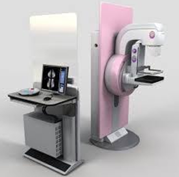
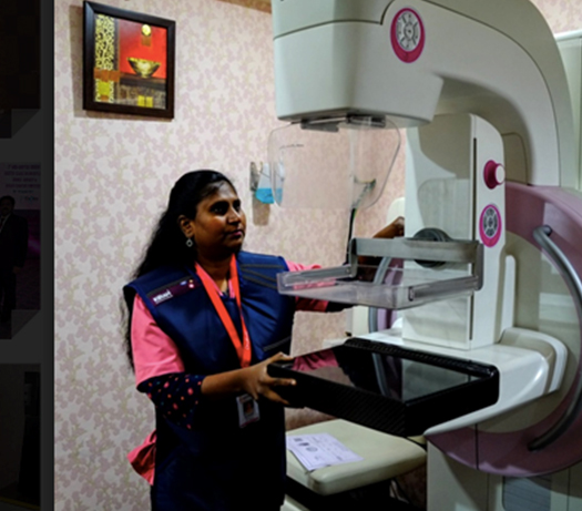
After clinical examination by the breast surgeon, patient (>40 yrs) is subjected to mammography in our clinic- Digital mammography with 3D tomosynthesis. It detects Stage 0 of Breast Cancer and is particularly used for detecting cancer in Indian women, since they have dense breasts. It has a plastic screen instead of a metal one, making the procedure painless. It acquires a series of images in 90+ slices, making the procedure accurate and quick, taking only 3-4 seconds. Screening with mammography has resulted in 15-20% reduction in mortality due to breast cancer. Sensitivity of mammography is 68% & specificity is 75%.
Interventions:
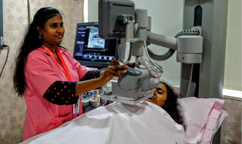
Ultrasound is an imaging test by which images of your breast can be viewed on a screen for detecting cancer, by sending high-frequency sound waves through it. It is the poor man’s MRI. At Orchid’s, we use ultrasound in combination with mammography, to increase the sensitivity of the test to nearly 90-95%.
High-density breasts make it difficult to detect tumour in these breasts using a mammogram, as both the dense tissue and tumour appear white. This is a common occurrence in India and therefore, at Orchids, we use the Automated Breast Volume Scanner (ABVS).
Why this technique?
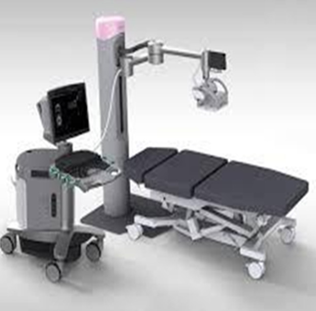
AUTOMATED BREAST VOLUME SCANNER AT ORCHIDS
AUTOMATED BREAST VOLUME SCANNER AT ORCHIDS
After clinical examination by the breast surgeon, patient (>40 yrs) is subjected to mammography in our clinic- Digital mammography with 3D tomosynthesis (machine name). Screening with mammography has resulted in 15-20% reduction in mortality due to breast cancer. Sensitivity of mammography is 68% & specificity is 75%.
Interventions
During breast cancer, the elastic properties of the affected tissues are altered. Elastography is a new technique that detects this change in elasticity and maps it by imaging. It is non-invasive, so the patient need not worry about getting any scars. It is used for characterization of difficult areas. But, doesn’t sonography detect changes in the breast as well? Why should one opt for elastography instead?
Elastography has a major advantage over sonography. Better resolution and clarity result in accurate detections. Whereas, sonography, alone, has the following limitations:
Benefits of elastography: